Amplifying the Voice of Clinical Neuroscience Medical Student Neurosurgery Training Center |
|

 What does the future of neuroimaging look like? By Lane Fitzsimmons
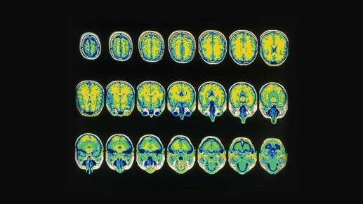
The integration of information science has the power to completely transform the medical field. The adoption of electronic health records supplies as much as 80 megabytes of clinical data per patient annually. (Huesch and Mosher) This wealth of information ripe for analysis provides the opportunity to highlight systemic shortcomings and shape a more efficient and data driven future for health care. When artificial intelligence tools perform tasks otherwise requiring human discernment, they can save valuable time, increase treatment accuracy, and improve patient outcomes. As one of the richest sources of patient data, medical imaging is the most promising immediate target of artificial intelligence applications. Carefully and quantitatively inspecting each and every megapixel of an X-ray, MRI, or CAT scan is a difficult and tedious task for radiologists. On average, clinicians take 13 minutes to read an MRI, while a machine learning model can complete the same task in four seconds. That’s 186 times faster! (American Heart Association) Not only are computers able to rapidly analyze large quantities of high resolution data, their pattern recognition is better than that of humans. (Basch 20-21) Algorithm readings are more capable of noticing nuanced details, as well as learning and improving as training data is provided. This enables them to recognize and make decisions upon patterns that humans do not yet fully understand. (Reardon) Current brain mapping research for poorly understood diseases employs artificial intelligence. By training algorithms on neuroimages of patients with mental illnesses, like depression and schizophrenia, researchers are elucidating the complex wirings and functions of these diseases (Marquez and Walid et al.). Algorithms work to consolidate enormous quantities of data from many sources into a comprehensive framework. In doing this, they enable researchers to determine which regions and connections are relevant to each mental illness. Identifying important indicative imaging characteristics would not only help clinicians better understand diseases, but also streamline diagnostic processes by automatically sorting and flagging suspect images. (Bresnick) Artificial neural networks, one of the most frequently utilized artificial intelligence algorithms, are doubly relevant to the field of neuroscience. Not only can these computing systems be relied upon to read and evaluate neuroimages, their framework aims to loosely model the cognitive processes of a biological neural network. Artificial neural networks were created to make decisions in a manner that imitates the connections of the human brain. Their structure relies on a network of nodes that pass signals along to each other just as actual biological neurons would. Each node has a weight threshold, only becoming activated and passing the signal to its neighbor once its individual output exceeds that value. From training data, a neural network learns to adjust the weights of different variables to maximize output accuracy. Complicated computational advancements, including training from output to input instead of input to output (backpropagation) and adding more and more layers of nodes (deep learning), have contributed to increased accuracy. (IBM Cloud) It is no surprise that sophisticated models like these rival human precision. Artificial intelligence models can also serve as neural surgical adjuncts and can even be used in real time. The sheer speed of algorithmic image analysis facilitates impactful intraoperative brain and spine imaging. Instead of relying solely upon preoperative scans, automated analysis enables surgeons to use updated MRIs and CTs. Analysis of these images in real time allows more accurate positioning for neuronavigation. (Li) Artificial intelligence imaging applications also serve a broad range of neurosurgical functions, such as planning the operative trajectory, indicating optimal positioning of microscopic cables, and alerting surgeons if they dissect too near high risk regions. (Panesar et al.) The FDA approves at least one new medical imaging algorithm per month, with the potential for broad neuroimaging applications. (Reardon) An automated classification model of 19 brain diseases demonstrated a 91% accuracy, faster than the average academic neuroradiologist. (Zaharchuk) Machine learning algorithms are utilized to differentiate MRI images of Alzheimer's patients from that of vascular dementia patients. (Castellazzi et al.) MRI-reading decision support algorithms have been created to do everything from determining the best time window to dissolve a blood clot to predicting the progression of Parkinson’s disease. (Lee et al. and Shu et al. ). With an estimated 84% of U.S. radiology clinics that have adopted or plan to adopt artificial intelligence programs, applications like these will be instrumental to the further algorithm development. Though the immediate predictive accuracy makes the potential for artificial intelligence apparent, clinicians should be hesitant to blindly rely upon uninterpretable algorithmic decision-making. “Back box” algorithms that boast high predictive accuracy often leave ample room for misunderstanding. For example, algorithms have been found to take into account correlations that are truly not indicative—such as time of day or clinic location. (Reardon) For linear models, it’s simple to access the role that each feature plays in a decision. This feature analysis is much more difficult with artificial intelligence algorithms, especially when models like neural networks are further mystified by complexities like back-propagation. It’s clear that only models that are rigorously trained and prospectively evaluated on diverse datasets are viable options for clinical application. Well-intentioned research can fall short, however, as large and diverse data sets are simply difficult to access while navigating privacy concerns. Even more, the “frame problem” suggests it is near impossible to account for the breadth of unpredictable situations an algorithm might face, like unknown objects on a scan. (Panesar et al.) The training and testing of aforementioned models with high output accuracy are almost exclusively conducted retrospectively, often on image sets with specific criteria and limited variability. Though research like this is essential to progress toward the creation of algorithms that are more robust, techniques are often not refined enough for clinical application. The high performance accuracy and potential utility of artificial intelligence makes future neuroimaging applications undoubtedly promising. There is, however, a long way to go before these strategies can be relied upon independently. Rather, artificial intelligence tools for neuroimaging will supplement, rather than replace, physician decision making. With that, artificial intelligence will play an increasingly important role in the future of medicine, but a computer won’t replace your doctor anytime soon. Works Cited: American Heart Association. “Machine learning could offer faster, more precise results for cardiac MRI scans.” 24 September 2019. Bash, Suzie. “Enhancing Neuroimaging with Artificial Intelligence.” Applied Radiology, vol. 49, no. 1, 2020, pp. 20-21, https://appliedradiology.com/communities/Artificial-Intelligence/enhancing-neuroimaging-with-artificial-intelligence. Bresnick, Jennifer. “Top 5 Use Cases for Artificial Intelligence in Medical Imaging.” Health IT Analytics, 30 October 2018, https://healthitanalytics.com/news/top-5-use-cases-for-artificial-intelligence-in-medical-i maging. Castellazzi, Gloria, et al. “A Machine Learning Approach for the Differential Diagnosis of Alzheimer and Vascular Dementia Fed by MRI Selected Features.” Frontiers in Neuroinformatics, 2020, https://www.frontiersin.org/articles/10.3389/fninf.2020.00025/full. Huesch, Marco D., and Timothy T. Mosher. “Using it or losing it? The case for data scientists inside health care.” Open Health News, 4 May 2017, https://www.openhealthnews.com/news-clipping/2017-05-04/using-it-or-losing-it-case-da ta-scientists-inside-health-care. IBM Cloud. “Neural Networks.” IBM Learn Hub, IBM, https://www.ibm.com/cloud/learn/neural-networks. Lee, Hyuanna, et al. “Machine Learning Approach to Identify Stroke Within 4.5 Hours.” Stroke, https://www.ahajournals.org/doi/10.1161/STROKEAHA.119.027611. Li, Qinghang. “Computer-assisted neurosurgery: Yesterday, today and tomorrow.” Journal of Neurology and Clinical Neuroscience, 2017. PULSUS, https://www.pulsus.com/scholarly-articles/computerassisted-neurosurgery-yesterday-toda y-and-tomorrow-3554.html. Marquez, Jennifer R. “Georgia State Researchers Developing Deep Learning Framework to Map Brain Disorders.” Georgia State News Hub, Georgia State University, 26 August 2020, https://news.gsu.edu/2020/08/26/georgia-state-researchers-developing-deep-learning-fra mework-to-map-brain-disorders/. Panesar, Sandip D., et al. “Promises and Perils of Artificial Intelligence in Neurosurgery.” Neurosurgery, vol. 87, no. 1, 2020, pp. 33-34, https://academic.oup.com/neurosurgery/article/87/1/33/5637041. Reardon, Sara. “Rise of Robot Radiologists.” Nature, 2019, https://www.nature.com/articles/d41586-019-03847-z. Shu, Zhen‐Yu, et al. “Predicting the progression of Parkinson's disease using conventional MRI and machine learning: An application of radiomic biomarkers in whole‐brain white matter.” Magnetic Resonance in Medicine, vol. 85, no. 3, 2021, pp. 1611-1624. Wiley Online Library, https://onlinelibrary.wiley.com/doi/abs/10.1002/mrm.28522. Walid, Yassin, et al. Machine-learning classification using neuroimaging data in schizophrenia, autism, ultra-high risk and first-episode psychosis, vol. 10, no. 278, 2020, https://www.nature.com/articles/s41398-020-00965-5. Zaharchuk, Greg Z. “Fellow in a Box: Combining AI and Domain Knowledge with Bayesian Networks for Differential Diagnosis in Neuroimaging.” Radiology, RNSA, 7 April 2020, https://pubs-rsna-org.proxy.library.cornell.edu/doi/full/10.1148/radiol.2020200819. Comments are closed.
|

3/11/2021